
USC Core Center of Excellence in Nano Imaging
Already have a Logon Account?
Main Contact Info
Emily Clough
USC Core Center of Excellence in Nano Imaging
1002 West Childs Way, MCB, Room LL109G
Los Angeles, CA 90015-9502
Remittance Contact Info
Emily Clough
USC- ATTN Emily Clough (CNI)
1150 S Olive St., Suite 1700
Los Angeles, CA 90015-9502
This facility has not published any Products. Please check back.
The following Products and Services are available within our facility:
Electron Microscope
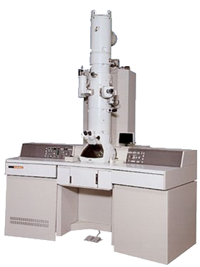 |
TEM JEOL 2100FJEOL 2100FThis JEOL 2100F is a transmission electron microscope equipped with a high resolution pole-piece (HRP) and thermal field emission source. The point-to-point resolution is 230 pm in TEM mode and 160 pm in STEM mode. TEM Images can be acquired on either the Orius Pre-GIF camera or the Gatan UltraScan 1000 Post-GIF camera. STEM images can be acquired using a JEOL BF detector or the GIF's BF/ADF detectors. In addition to high resolution imaging, the microscope is equipped for both electron energy loss spectroscopy (Quantum SE GIF) and x-ray spectroscopy (Oxford X-MaxN 100 TLE Windowless SDD). Both single tilt (+/- 40 Degrees) and double |
|
|
TEM FEI Talos F200CFEI Talos F200C |
|
|
SEM JEOL 7001FJEOL 7001FThe JSM-7001F-LV is an analytical field emission scanning electron microscope equipped with imaging detectors for secondary and backscattered electrons it has analytical detectors for electron backscatter diffraction (EBSD) and energy dispersive X-ray spectrometer (EDS). The low vacuum mode can be used for imaging of untreated non-conductive specimens. |
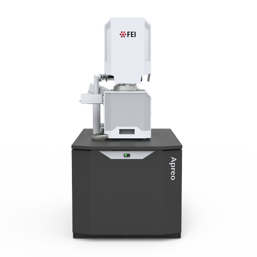 |
SEM FEI Nova NanoSEM 450FEI Nova NanoSEM 450Field emission SEM with ultra-stable, high current Schottky gun, Advanced optics and detection, including an immersion mode, beam deceleration, in-lens TLD-SE and -BSE, DBS and STEM for best selection of the information and image optimization |
|
|
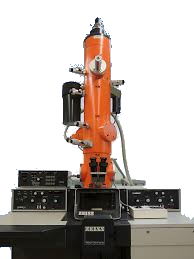
FIB JEOL 4500JEOL 4500The JIB-4500 is a high performance LaB6 SEM with Ga liquid metal ion source FIB. It is equipped with EDAX energy dispersive X-ray spectrometer (EDS) and Omniprobe AutoProbe 200 in situ TEM lift-out and Omniprobe OmniGIS gas injector system.. |
Photon Microscope
Super Resolution GE DeltaVisonGE DeltaVisonThe GE DeltaVision OMX system is an optical-sectioning epifluorescence microscope capable of operating in 3D-SIM, TIRF or widefield-deconvolution mode with the option of stage/sample heating, CO2 capabilities and live-cell observations. In most applications sample preparation/engineering is "nearly" identical to conventional light microscopy. Excitation lines include 405, 445, 488, 514, 568, 642nm; multiplexing up to 4 colors is possible. 3 sCMOS cameras provide good sensitivity with near simultaneous acquisition. Speed in 3D-SIM mode is enhanced by the "Blaze" light path which manipulates the structured illumination pattern without |
Ion Microscope
 |
HIM Zeiss OrionGE DeltaVisonThe ORION NanoFab uses a ion beam to achieve imaging with both conductive and non-conductive samples with the use of a electron flood gun for charge compensation. A higher depth of field compared to images acquired with FE-SEMs and greater surface sensitivity. A gas injection system (GIS) with the Fibics Nanopatterning and Visualization Engine (NPVE) is attached for lithography. |
Spectrometer
XPS Kratos Axis UltraKratos Axis UltraThe Kratos Axis Ultra DLD is an X-ray photoelectron spectrometer (XPS) equipped with a magnetic immersion lens and charge neutralisation system with the spherical mirror and concentric hemispherical analyzers with the choice of either a Mg or Al anodes for high-sensitivity surface analysis. The instrument has a typical sampling depth of 2-10 nm and detection limit around 1 ppt (varies depending on element of interest). The hemispherical analyzer provides both high energy resolution and high sensitivity spectroscopic performance. The small spot capabilities of less than 15 microns are achieved via a series of selected area apertures |
|
XRD Bruker D8Bruker D8The D8 ADVANCE is an all-purpose X-ray analyzer which can be configured for all powder diffraction applications, including phase identification, quantitative phase analysis, micro-structure and crystal structure analysis, residual stress and texture investigations, X-ray reflectometry, and micro-diffraction, depending on accessories. |
|
XRF Bruker S8Bruker S8The S8 TIGER is a high performance wavelength-dispersive X-ray fluorescence spectrometer (WDXRF) suitable for fast qualitative, quantitative, and “standardless” analysis of elemental composition from oxygen to uranium. Standardless analysis (QuantExpress) is capable of up to 0.1% accuracy, whereas a calibrated sample matrix can attain < 1 ppm sensitivity. The 4kW S8 uses a rhodium X-ray tube, scintillation and proportional flow detectors, and is capable of analyzing fluorescence energies from 0-100 keV. It also uses a robotic sample changer to allow for high-throughput of many samples. The S8 is equipped with 3 crystals (LiF200, X |
Sample Preparation
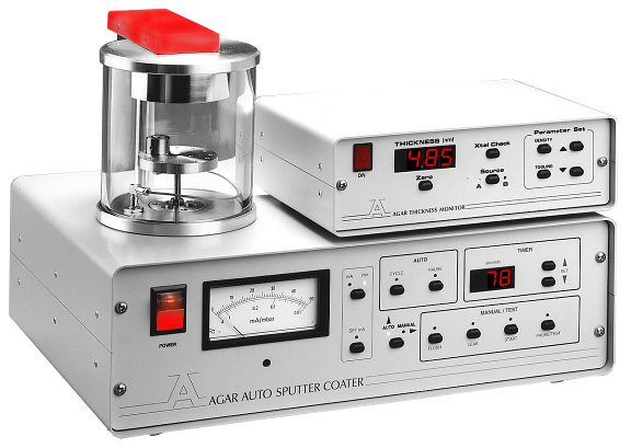 |
Cressington 108 Manual Sputter CoaterCressington 108 Manual Sputter CoaterSample coating with Au, Pd or Pt for non-conducting samples for standard SEM imaging. |
|
|
Cressington 108C Auto Carbon CoaterCressington 108C Auto Carbon CoaterCarbon coating for non-conductive specimens with a rod evaporation system giving a thickness of around 20nm for SEM imaging and x-ray micro-analysis (EDS). |
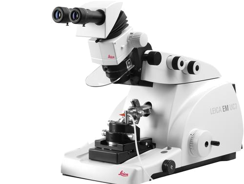
Leica EM UC6 Cryo-UltramicrotomeLeica EM UC6 Cryo-UltramicrotomeLeica Ultramicrotome can be used to freeze thin films for sectioning. Suitable for all soft materials (biological, polymers, etc.) |
|
 |
Reichert Jung UltracutEReichert Jung UltracutEUsed for cutting specimens into ultra-thin sections for the TEM. |
Tousimis 815 Critical Point DryerTousimis 815 Critical Point DryerDehydrating biological specimens prior to examination in the Scanning Electron Microscope. |
|
XEI CombiClean Plasma CleanerXEI CombiClean Plasma CleanerCleans SEM/TEM samples and TEM holders with an energy efficient capacitive coupled plasma. |
This facility has not published any News. Please check back.
Quick Quotes have not been configured. Please check back soon (Code 001, Code 002)
, please enter that email address here.")